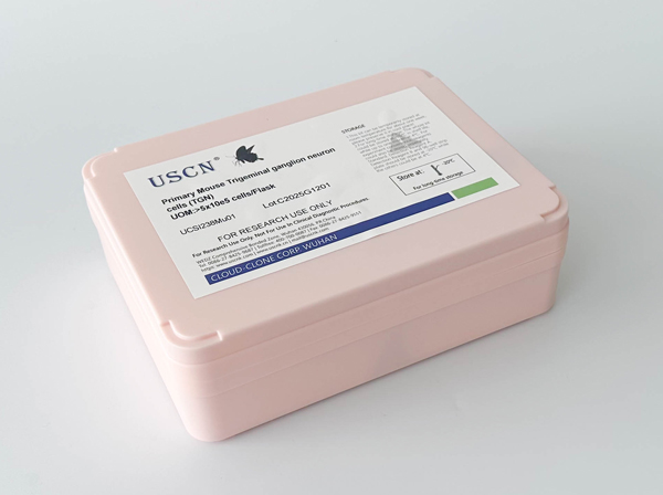
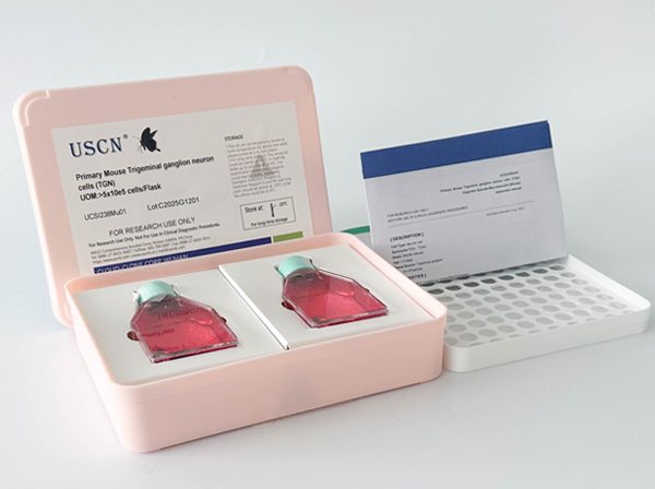
Primary Canine Neonatal Epidermal Keratinocytes (NEK)
Neonatal Epidermal Keratinocyte Cells
- Product No.UCSI075Ca01
- Organism SpeciesCanis familiaris; Canine (Dog) Same name, Different species.
- Tissue SourceNeonatal Epidermal tissue
- Growth PropertiesAdherent
- Cell TypeEpidermal Keratinocytes
- DiseaseNormal
- ApplicationsPrimary Cells
- Download Instruction Manual
- UOM 5X10^5
-
FOB
US$ 690
For more details, please contact local distributors!
Biosafety of the Primary Canine Neonatal Epidermal Keratinocytes (NEK)
Primary Canine Neonatal Epidermal Keratinocytes (NEK) are negative for HIV-1, HBV, HCV, mycoplasma, bacteria, yeast and fungi.
Cluture Conditions of the Primary Canine Neonatal Epidermal Keratinocytes (NEK)
Keratinocyte-SFM+ 1% Keratinocyte Growth Supplement +1%Penicillin-Streptomycin Solution. Temperature: 37°C. Condition: 95% air, 5% carbon dioxide
Product Format of the Primary Canine Neonatal Epidermal Keratinocytes (NEK)
Frozen 1 mL or T25 flask.
USAGE of the Primary Canine Neonatal Epidermal Keratinocytes (NEK)
Upon receiving the cells in a T-25 flask at room temperature, immediately transfer the cells to 37°C, 5% incubator; the cells in vials, directly and immediately transfer the cells from dry ice to liquid nitrogen.
GIVEAWAYS
INCREMENT SERVICES
Lysis Buffer Specific for ELISA / CLIA
Staining Solution for Cells and Tissue
Primary Cells Customized Service
Total Protein/DNA/RNA Extract Customized Service
Cell Culture Medium Customized Service
Immunocytochemistry (ICC) Experiment Service
Flow Cytometry (FCM) Experiment Service
Cell Function Experiment Service
In Situ Hybridization (ISH) Experiment Service
Transmission Electron Microscopy (TEM) Experiment Service
Scanning Electron Microscope (SEM) Experiment Service
MTT Cell Proliferation and Cytotoxicity Assay Kit
Cell Invasion Assay Service
Cell Wound Scratch Assay Service
Cell Proliferation / Toxicity Assay Service
Cell Colony Formation Assay Service
Cell Migration Assay Service
CCK-8 Kit
Serum-free 293 cell medium
HE Stain
Nitric Oxide Assay Kit (A012)
Nitric Oxide Assay Kit (A013-2)
Total Anti-Oxidative Capability Assay Kit(A015-2)
Total Anti-Oxidative Capability Assay Kit (A015-1)
Superoxide Dismutase Assay Kit
Fructose Assay Kit (A085)
Citric Acid Assay Kit (A128 )
Catalase Assay Kit
Malondialdehyde Assay Kit
Glutathione S-Transferase Assay Kit
Microscale Reduced Glutathione assay kit
Glutathione Reductase Activity Coefficient Assay Kit
Angiotensin Converting Enzyme Kit
Glutathione Peroxidase (GSH-PX) Assay Kit
Cloud-Clone Multiplex assay kits
Related products
| Catalog No. | Organism species: Canis familiaris; Canine (Dog) | Applications (RESEARCH USE ONLY!) |
| UCSI075Ca01 | Primary Canine Neonatal Epidermal Keratinocytes (NEK) | Primary Cells |
| UMSI075Ca11 | Medium for Canine Neonatal Epidermal Keratinocytes (NEK) | Medium for Canine Neonatal Epidermal Keratinocytes |


