
Active Tumor Necrosis Factor Receptor 1 (TNFR1)
CD120A; P55; TNFRSF1A; TBP1; FPF; TNF-R; TNF-R-I; TNF-R55; TNFAR; TNFR55; TNFR60; P55-R; P60; Tumor necrosis factor receptor 1; Tumor necrosis factor-binding protein 1
- Product No.UAPB499Hu01
- Organism SpeciesHomo sapiens (Human) Same name, Different species.
- Buffer Formulation20mM Tris, 150mM NaCl, pH8.0, containing 1mM EDTA, 1mM DTT, 0.01% SKL, 5% Trehalose and Proclin300.
- TraitsFreeze-dried powder
- Purity> 95%
- Isoelectric Point6.4
- ApplicationsCell culture; Activity Assays.
- Download Instruction Manual
- UOM 10µg50µg 200µg 1mg 5mg
-
FOB
US$ 112
For more details, please contact local distributors!US$ 279
For more details, please contact local distributors! US$ 558
For more details, please contact local distributors! US$ 1674
For more details, please contact local distributors! US$ 4185
For more details, please contact local distributors!
-
 Packages (Simulation)
-
 Packages (Simulation)
-
 Figure. SDS-PAGE
-
Figure. Western Blot
-
ISO9001: 2008, ISO13485: 2003 Registered
ACTIVITY TEST of the Active Tumor Necrosis Factor Receptor 1 (TNFR1)
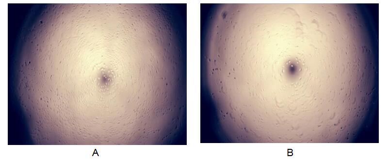
Figure. The apoptosis of A549 cells by TNFα was inhibited by TNFRSF1B.
Tumor necrosis factor receptor superfamily member 1B (TNFRSF1B), also known as tumor necrosis factor receptor 2 (TNFR2) and CD120b, is a membrane receptor that binds tumor necrosis factor-alpha (TNFα). This protein and TNF-receptor 1 form a heterocomplex that mediates the recruitment of two anti-apoptotic proteins, c-IAP1 and c-IAP2, which possess E3 ubiquitin ligase activity. TNFRSF1B can inhibit cell apoptosis which induced by TNFα. Briefly, A549 cells were seeded into triplicate wells of 96-well plates at a density of 2,000 cells/well and allowed to attach, replaced with serum-free overnight, then the medium was replaced with 2% serum standard DMEM including 1μg/mL TNFα and various concentrations of recombinant human TNFRSF1B. After incubated for 96h, cells were observed by inverted microscope and cell proliferation was measured by Cell Counting Kit-8 (CCK-8). Briefly, 10µL of CCK-8 solution was added to each well of the plate, then the absorbance at 450nm was measured using a microplate reader after incubating the plate at 37°C for 1-4 hours. Apoptosis of A549 cells had been inhibit after incubation with TNFRSF1B for 96h observed by inverted microscope was shown in Figure 1. Cell viability was assessed by CCK-8 (Cell Counting Kit-8) assay after incubation with recombinant TNFRSF1B for 96h. The result was shown in Figure 2. It was obvious that TNFRSF1B significantly suppress cell apoptosis induced by TNFα.
(A) A549 cells cultured in DMEM contain 1μg/mL TNFα and 0.1ng/mL TNFRSF1B for 96h;
(B) A549 cells cultured in DMEM only contain 1μg/mL TNFα for 96h.
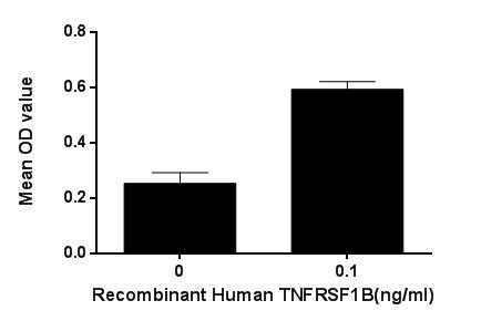
Figure. TNFRSF1B suppress the apoptosis of A549 cells induced by TNFα.
USAGE of the Active Tumor Necrosis Factor Receptor 1 (TNFR1)
Reconstitute in 20mM Tris, 150mM NaCl (PH8.0) to a concentration of 0.1-1.0 mg/mL. Do not vortex.
STORAGE of the Active Tumor Necrosis Factor Receptor 1 (TNFR1)
Avoid repeated freeze/thaw cycles. Store at 2-8°C for one month. Aliquot and store at -80°C for 12 months.
STABILITY of the Active Tumor Necrosis Factor Receptor 1 (TNFR1)
The thermal stability is described by the loss rate. The loss rate was determined by accelerated thermal degradation test, that is, incubate the protein at 37°C for 48h, and no obvious degradation and precipitation were observed. The loss rate is less than 5% within the expiration date under appropriate storage condition.
INCREMENT SERVICES
BCA Protein Quantification Kit
Molecular Mass Marker for Protein
Monoclonal Antibody Customized Service
Polyclonal Antibody Customized Service
Protein Activity Test Experiment Service
Electrophoretic Mobility Shift Assay (EMSA) Experiment Service
Buffer
Lentivirus Packaging Experiment Service
Adenovirus Packaging Experiment Service
Real Time PCR Experimental Service
Spike RBD Protein (S-RBD)
Protein G
Protein A