
Active Programmed Cell Death Protein 1 (PD1)
CD279; PDCD1; SLEB2; HPD1P
- Product No.UAPA751Hu02
- Organism SpeciesHomo sapiens (Human) Same name, Different species.
- Buffer FormulationPBS, pH7.4, containing 0.01% SKL, 5% Trehalose.
- TraitsFreeze-dried powder
- Purity> 90%
- Isoelectric Point8.7
- ApplicationsCell culture; Activity Assays.
- Download Instruction Manual
- UOM 10µg50µg 200µg 1mg 5mg
-
FOB
US$ 70
For more details, please contact local distributors!US$ 175
For more details, please contact local distributors! US$ 350
For more details, please contact local distributors! US$ 1050
For more details, please contact local distributors! US$ 2625
For more details, please contact local distributors!
-
 Packages (Simulation)
-
 Packages (Simulation)
-
 Figure. SDS-PAGE
-
ISO9001: 2008, ISO13485: 2003 Registered
ACTIVITY TEST of the Active Programmed Cell Death Protein 1 (PD1)
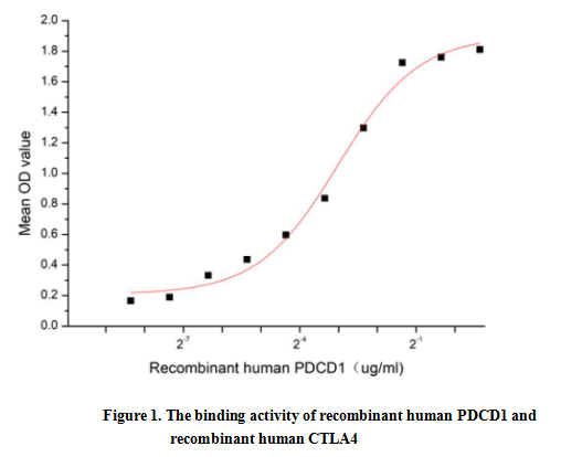
PD-1 (Programmed Death-1 receptor), also known as CD279, is a receptor expressed on T cells responsible for modulating T cell activation. Like CTLA 4, PD-1 is classified as an immune checkpoint inhibitory receptor. When PD-1 protein binds to PD-L1, it initiates a negative signaling cascade inhibiting activation of T cells. The cytoplasmic tail contains two tyrosine residues that form the immuno receptor tyrosine-based inhibitory motif (ITIM) and immuno receptor tyrosine-based switch motif (ITSM) that are important for mediating PD-1 signaling. Normally, PD-1 helps keep T cells from attacking other cells in the body. However, many cancers take advantage of this by expressing high amounts of PD-L1 allowing cancer cells to evade the body's own immune response. Blocking the PD-1:PD-L1 interaction has proven successful in treating many different cancer types. A functional binding ELISA assay was conducted to detect the interaction of recombinant human PDCD1 and recombinant human CTLA4. Briefly, PDCD1 was diluted serially in PBS with 0.01% BSA (pH 7.4). Duplicate samples of 100 μl were then transferred to CTLA4-coated microtiter wells and incubated for 1h at 37℃. Wells were washed with PBST and incubated for 1h with anti-PDCD1 pAb, then aspirated and washed 3 times. After incubation with HRP labelled secondary antibody for 1h at 37℃, wells were aspirated and washed 5 times. With the addition of substrate solution, wells were incubated 15-25 minutes at 37℃. Finally, add 50 µL stop solution to the wells and read at 450/630 nm immediately. The binding activity of recombinant human PDCD1 and recombinant human CTLA4 was shown in Figure 1, the EC50 for this effect is 0.12 ug/mL.
USAGE of the Active Programmed Cell Death Protein 1 (PD1)
Reconstitute in 10mM PBS (pH7.4) to a concentration of 0.1-1.0 mg/mL. Do not vortex.
STORAGE of the Active Programmed Cell Death Protein 1 (PD1)
Avoid repeated freeze/thaw cycles. Store at 2-8°C for one month. Aliquot and store at -80°C for 12 months.
STABILITY of the Active Programmed Cell Death Protein 1 (PD1)
The thermal stability is described by the loss rate. The loss rate was determined by accelerated thermal degradation test, that is, incubate the protein at 37°C for 48h, and no obvious degradation and precipitation were observed. The loss rate is less than 5% within the expiration date under appropriate storage condition.
INCREMENT SERVICES
BCA Protein Quantification Kit
Molecular Mass Marker for Protein
Monoclonal Antibody Customized Service
Polyclonal Antibody Customized Service
Protein Activity Test Experiment Service
Electrophoretic Mobility Shift Assay (EMSA) Experiment Service
Buffer
Lentivirus Packaging Experiment Service
Adenovirus Packaging Experiment Service
Real Time PCR Experimental Service
Spike RBD Protein (S-RBD)
Protein G
Protein A